
Figure 2.
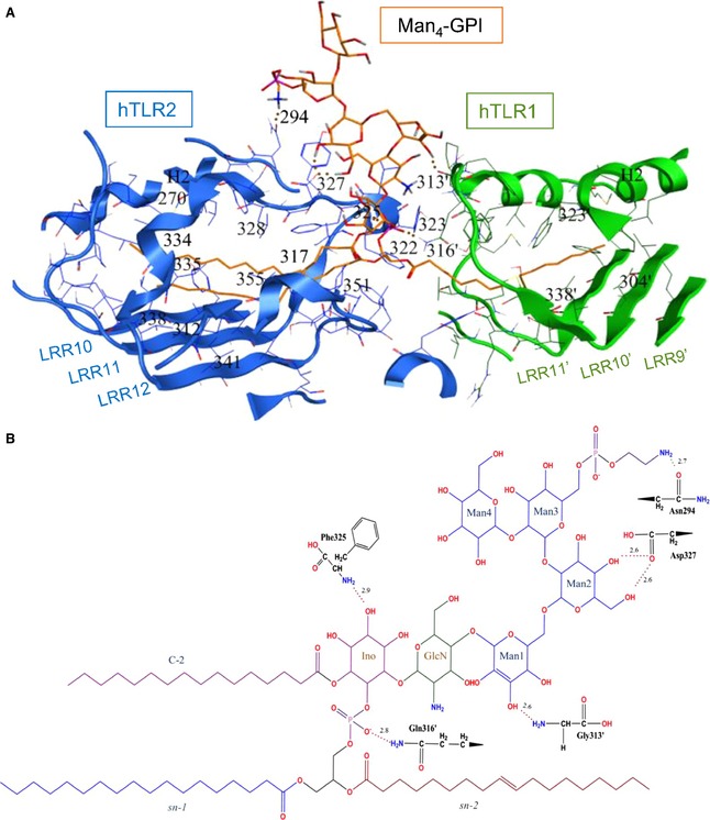
The Man4‐GPI binding site in the docked structure of the hTLR2‐hTLR1 complex. (A) The hTLR2 and hTLR1 residues involved in Man4‐GPI binding are shown in blue and green, respectively. The hydrogen bonds are shown by dotted red lines. Carbons, nitrogens, oxygens and phosphorous of Man4‐GPI are coloured in orange, blue, red and purple, respectively. (B) The chemical structure of Man4‐GPI and residues forming hydrogen bonds. Hydrogen bonds are shown with dotted red lines, and their bond distances are written above the lines in angstroms. Carbons, nitrogens, oxygens and phosphorous of Man4‐GPI are coloured in black, blue, red and purple, respectively. Apostrophes are used for hTLR1 residues to differentiate from residues in hTLR2.