
Figure 4.
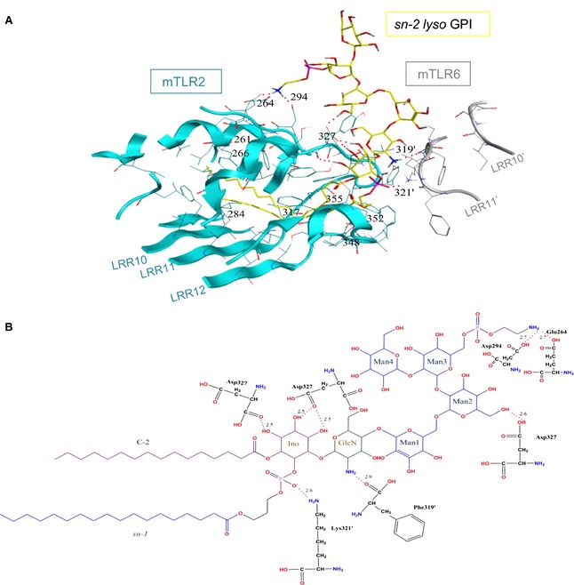
The sn‐2 lyso GPI binding site in the docked structure of the mTLR2‐mTLR6 complex. (A) The mTLR2 and mTLR6 residues involved in sn‐2 lyso GPI binding are shown in cyan and grey, respectively. The hydrogen bonds are shown by dotted red lines. Carbons, nitrogens, oxygens and phosphorous of the sn‐2 lyso GPI are coloured in yellow, blue, red and purple, respectively. (B) The chemical structure of sn‐2 lyso GPI and residues forming hydrogen bonds. Hydrogen bonds are shown with dotted red lines, and their bond distances are written above the lines in angstroms. Carbons, nitrogens, oxygens and phosphorous of sn‐2 lyso GPI are coloured in black, blue, red and purple, respectively. Apostrophes are used for mTLR6 residues to differentiate from residues in mTLR2. Residue Asp327 forms multiple hydrogen bonds with the ligand and so it is shown more than once.