
Figure 5.
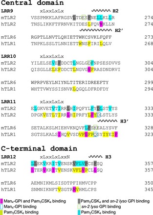
Structure‐based sequence alignment of the ligand‐binding regions of hTLR2, hTLR1, mTLR2 and mTLR6. The ligand binding regions of hTLR2, hTLR1, mTLR2 and mTLR6 sequences are aligned based on their structures. Conserved leucines and residues in the asparagine ladder are written above the sequences. The positions of α‐helices are indicated by coils above the sequences and are numbered.