Fig. 1.
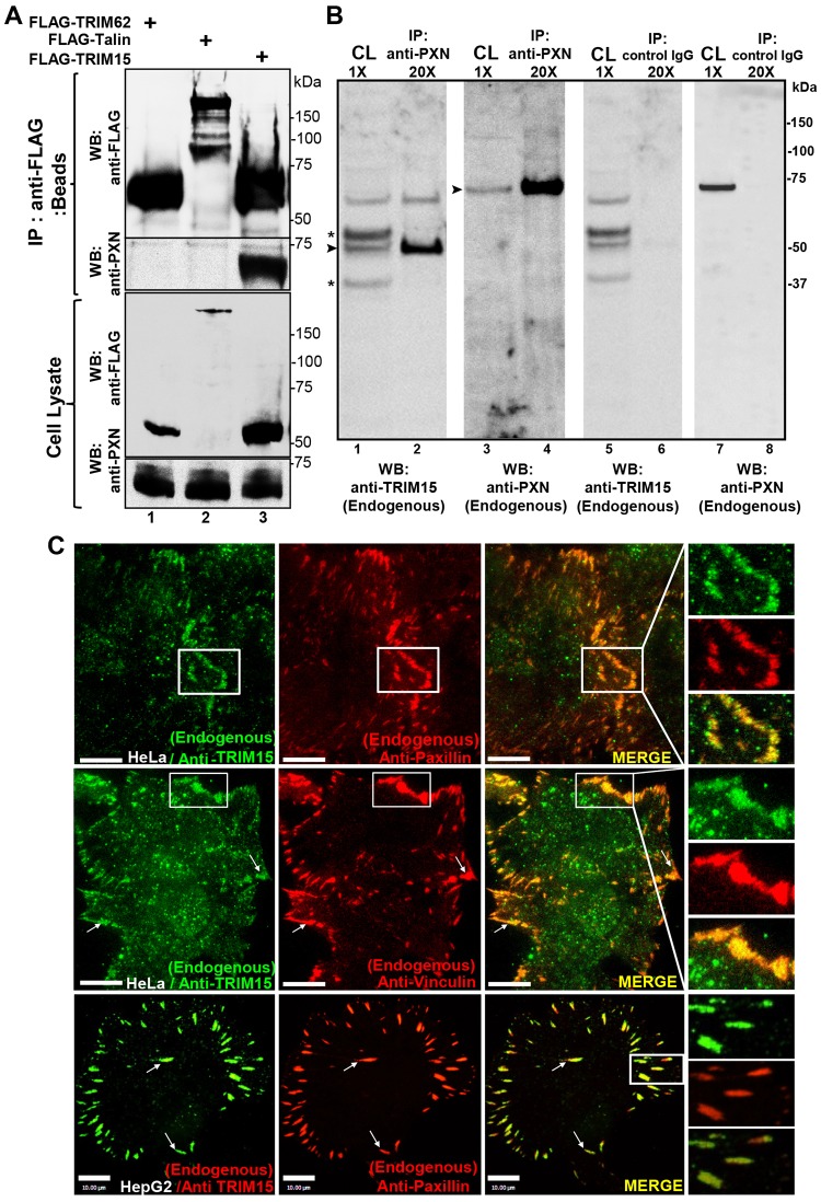
TRIM15 interacts with paxillin and localizes to focal adhesions. (A) Western blot (WB) analyses of anti-FLAG immunoprecipitates (beads) and cell lysates using antibodies against the FLAG epitope or paxillin (PXN) from HeLa cell lysates expressing FLAG-tagged TRIM15 or control proteins TRIM62 and talin. (B) Western blot analyses of anti-paxillin (endogenous) or control IgG immunoprecipitates (IP) and cell lysates (CL) with antibodies against endogenous TRIM15 or paxillin from HepG2 cell lysates. Asterisk, non-specific background bands; arrowheads, TRIM15- or paxillin-specific band. The relative concentrations of immunoprecipitates (20×) with respect to cell lysates (1×) loaded on the gel are also indicated. (C) Individual and merged TIRFM images of the indicated cell lines showing endogenous TRIM15, paxillin and vinculin detected by immunofluorescence using respective antibodies. Arrows point to colocalized signals in individual focal adhesions. Areas outlined in white are shown at a higher magnification to the right. Scale bars: 10 µm.

