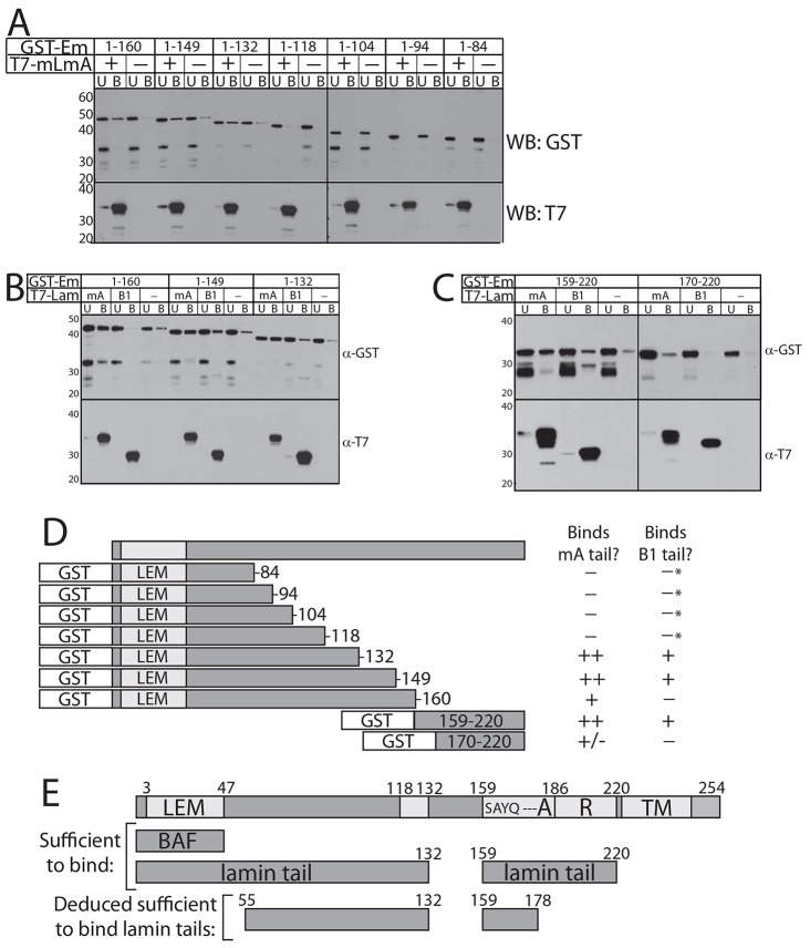
Fig. 3.
Lamin tails contact two regions of emerin. (A–C) Immunoblots of bound (B label, 25% loaded) vs unbound (U label; 4% loaded) fractions of each recombinant GST–emerin polypeptide (1.0 µM) incubated with NTA-agarose-immobilized recombinant lamin A or B1 tail baits, probed for GST (WB: GST), then stripped and reprobed for the lamin T7 tag (WB: T7). (A) Lamin bait: N-terminally T7-tagged and C-terminally His-tagged mature lamin A tail residues 385–646 (‘T7-mLmA’; n = 3). (B,C) Two baits were tested: mature lamin A tail (‘mA’; see A) or lamin B1 tail residues 395–586 (‘B1’; n = 3 each). (D) Schematic summary of results in A–C. The number of ‘+’ symbols indicates the degree of binding; ‘–’ indicates no binding; ‘+/–’ indicates inconsistent, weak binding. *Binding to lamin B1 not detected in two independent trials (our unpublished observations). (E) Emerin schematic indicating the LEM domain (LEM; sufficient to bind BAF), residues S159AYQ162, region ‘A’ (residues 168–186), region ‘R’ (residues 187–220), the transmembrane domain (TM) and two regions (residues 1–132 and 159–220) each sufficient to bind lamin A or B1 tails in vitro.

