
Fig. 8.
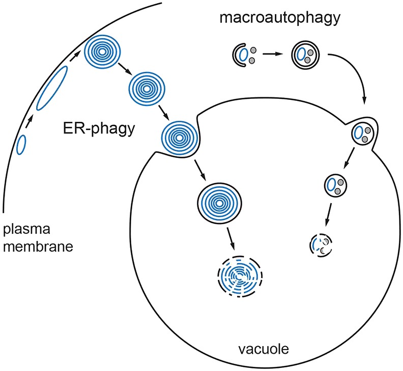
Model of the autophagic response to ER stress. ER stress induces expansion of the peripheral ER (blue) and formation of ER whorls, which are selectively taken up into the vacuole by ER-phagy. Concomitantly, macroautophagy is activated. Forming autophagosomes (crescent-shaped membrane sac) engulf pieces of the ER (blue) as well as other cytoplasmic constituents (gray). ER-phagy and macroautophagy might act independently and in parallel, as shown in this model, but could also be linked.