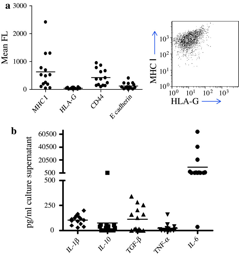
Fig. 1.
Primary ovarian cancer cells isolated from patients with progressive disease express high levels of MHC class I, low levels of HLA-G and high levels of immunosuppressive cytokines. Primary ovarian cancer cells were obtained from the ascitic fluid of 14 ovarian cancer patients and analysed by FACS for the presence of MHC class I, CD44, E-cadherin and HLA-G. a shows mean fluorescence for each sample, together with a representative FACS profile of tumour cells from AD patient 2 co-stained with MHC I and HLA-G. Primary tumour cells were seeded at 1 × 10⁶/ml, cultured in vitro for 48 h and supernatants collected for cytokine analysis. b shows the concentration of IL-1β, IL-10, TGF-β, TNF-α and IL-6 in pg/ml in cell culture supernatants as determined by ELISA. Four samples secreted more than 2,000 pg/ml IL-6 and are shown as 1,500 pg/ml

