
Fig. 7. Rab36 recruits JIP4 to recycling endosomes.
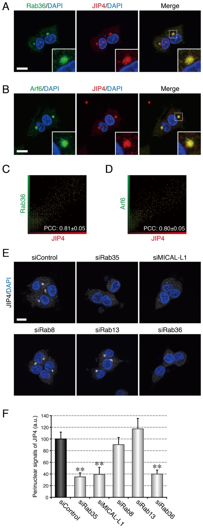
(A) Colocalization between Rab36 and JIP4 in PC12 cells. After NGF stimulation for 6 hr PC12 cells were fixed and stained with anti-Rab36 antibody, anti-JIP4 antibody, and DAPI. (B) Colocalization between Arf6 and JIP4 in PC12 cells. After NGF stimulation for 6 hr PC12 cells were fixed and stained with anti-Arf6 antibody, anti-JIP4 antibody, and DAPI. The insets in panels A and B are magnified views of the boxed areas. (C,D) Intensity scatter plot of Rab36 signals versus JIP4 signals (C) and of Arf6 signals versus JIP4 signals (D) in PC12 cells after NGF stimulation for 6 hr. The Pearson's correlation coefficient (PCC) value (mean ± SD) for the relation between them is shown at the bottom (n = 30 from 3 independent experiments). (E) Disappearance of JIP4 signals from the perinuclear area of Rab35-depleted, MICAL-L1-depleted, and Rab36-depleted PC12 cells. After NGF stimulation for 6 hr PC12 cells treated with siControl, siRab35, siMICAL-L1, siRab8, siRab13, or siRab36 were fixed and stained with anti-JIP4 antibody and DAPI. Scale bars: 10 µm. (F) Perinuclear JIP4 signals (mean and SE) of siControl-treated, siRab35-treated, siMICAL-L1-treated, siRab8-treated, siRab13-treated, and siRab36-treated PC12 cells after NGF stimulation for 6 hr (n = 60 from 3 independent experiments).