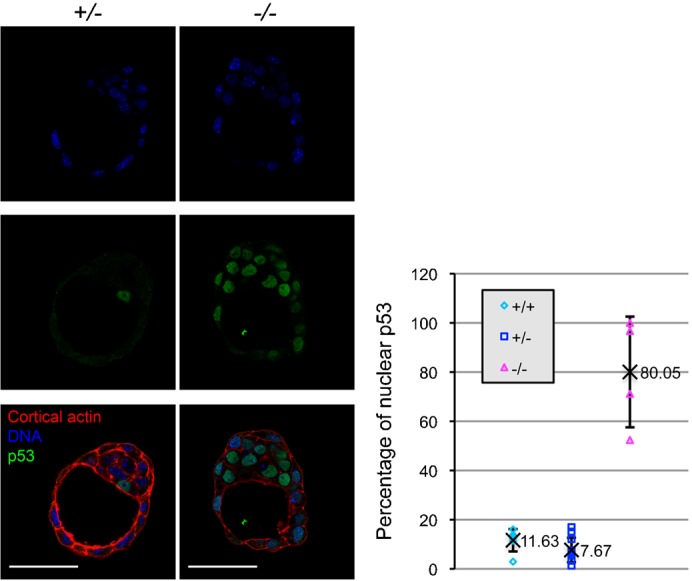
Fig. 5. PDCD2 knockout in early embryos induces nuclear p53.

Left panel: confocal sections of heterozygous (+/−) and homozygous (−/−) PDCD2 knockout 3.5 dpc embryos stained for DNA (Hoechst, blue), cortical actin (Phalloidin, red) and p53 (green); right panel: quantification of percentage nuclear p53 cells per embryo (TCN quantified based on Hoechst staining). Homozygous knockout embryos contain significantly more nuclear p53 cells (average 80%, N = 4) than WT (average 11.6%, N = 11, p = 7.E−5 by t test) and heterozygous embryos (average 7.7%, N = 4, p = 9.E−8 by t test). Scale bars: 50 µm.
