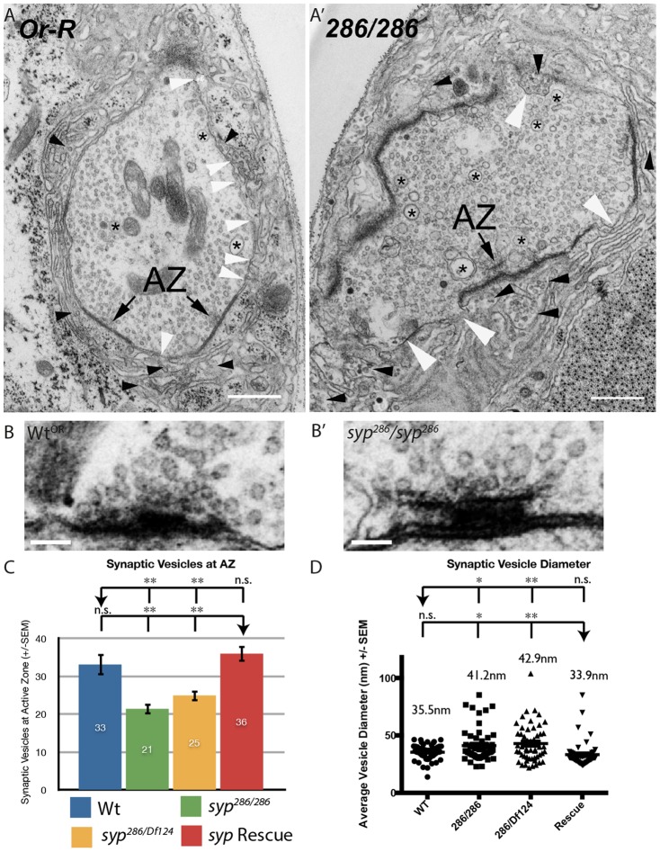
Fig. 2. Syncrip mutants exhibit defects in synapse structure and vesicle docking.
(A,A′) Ultrastructure analysis of syncrip mutant synapses reveals dramatic defects in synapse structure and organization. syp mutant boutons contain a larger population of large vesicles (asterisk) than wildtype and exhibit multiple points at which the pre- and postsynaptic membrane are not apposed (white arrowheads). While lesions are present in the wildtype boutons also, they are much more prevalent in syp mutants. In syp mutants large clusters of synaptic vesicles are present in the postsynapse (black arrow heads), though this likely is caused by fixation and sectioning and reflects a weakness in synaptic architecture relative to wildtype (see main text). (B–C) Consistent with a deficit in vesicle release, syp mutants have fewer vesicles docked, or close to, active zones relative to controls. Vesicles were counted within a 500 nm × 25 nm area around active zones in wt (n = 7), syp286/syp286 (n = 14), syp286/sypDf124 (n = 25), syp Rescue (n = 13). The wildtype T-bar was chosen as it is representative of the number of docked vesicles observed. syp mutant terminals contained enlarged T-bars. (D) syncrip mutants exhibit increased vesicle size (n>30 vesicles counted across ≥3 boutons for each test). Scale bars: (A,A′) 500 nm, (B,B′) 100 nm. Independent two-tailed Student's t-test; *** p<0.001 ** p<0.005 * p<0.05, n.s. p>0.05.

