
Fig. 3. Syncrip is enriched in muscle nuclei and at the postsynapse, but is undetectable at the presynapse.
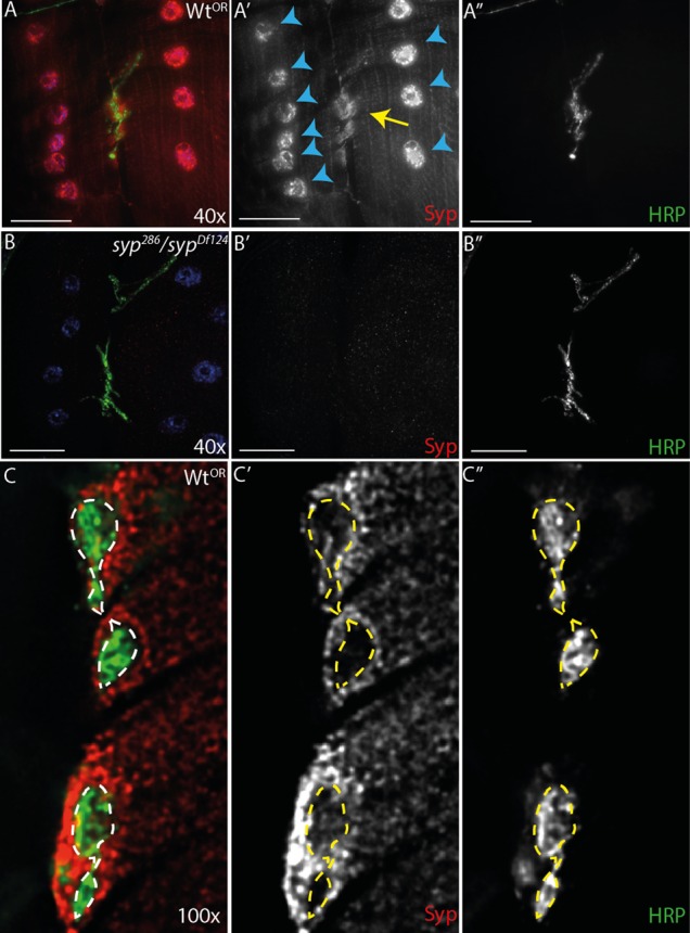
(A–A″) Sensitive wide-field imaging coupled with a polycolonal antibody reveals Syncrip throughout the muscle cytoplasm, with enrichment in the nuclei (blue arrowheads) and at the postsynapse (yellow arrow). (B–B″) The Syncrip antibody is highly specific and registers little signal in the syp mutants. (C–C″) Higher magnification imaging fails to robustly detect Syncrip in the presynapse above background fluorescence. Images are maximum intensity 5 µm projections. Scale bars: (A–B″) 40 µm, (C–C″) 5 µm.