
Figure 7.
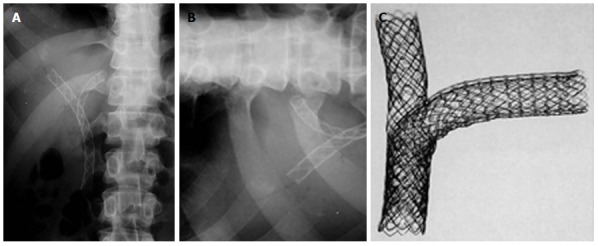
Percutaneous double stenting. A: X-ray showing the Y configuration of the stent (Boston Scientific MA, United States); B: Transverse limb of a T stent (Taewoong Medial, South Korea) showing the open mesh in the center; C: Assembly of a T stent showing the vertical stent passing through the open mesh of the transverse stent.