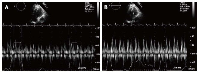
Figure 3.

Apical four chamber view of transthoracic echocardiogram and pulse wave Doppler’s recording. A: Tricuspid valve inflow in a patient with constrictive pericarditis; B: Mitral valve inflow in a patient with constrictive pericarditis. Note with inspiration, the E-wave velocity of mitral valve inflow decreases significantly. This reflects the hemodynamic changes in constrictive pericarditis, resulting from the lack of intrathoracic pressure transmission to the cardiac chambers and the exaggerated ventricular interdependence.
