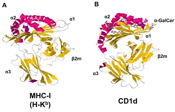
Fig. 1. The structures of MHC class I and CD1d. The side views of MHC-I (A) and CD1d (B) molecules. α1 and α2 domains consisting two alpha helixes on top of the molecules (magenta) and a beta-plated sheet (yellow) beneath the alpha helixes form an antigen binding region, which is facing toward T cell receptors. β2m-associated α3 domain is attached on the cell surface. PDB accession numbers are 2VAA (MHC I) and 1ZT4 (CD1d).

