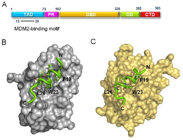
Fig. 2. Structural similarity between MDM2/p53TAD and Bcl-2/p53TAD complexes. (A) Domain organization of p53. p53 consists of a transactivation domain (TAD), proline-rich domain (PR), DNA-binding domain (DBD), oligomerization domain (OD), and C-terminal domain (CTD). The residues 15-29 of p53TAD are indicated as MDM2-binding motif. (B) Crystal structure of the MDM2/p53TAD peptide (residues 15-29) complex (PDB code: 1YCR) (9). (C) A refined structural model for the Bcl-2/p53TAD peptide (residues 15-29) complex generated from an NMR data-driven structure calculation (21). The p53TAD peptide is shown in green.