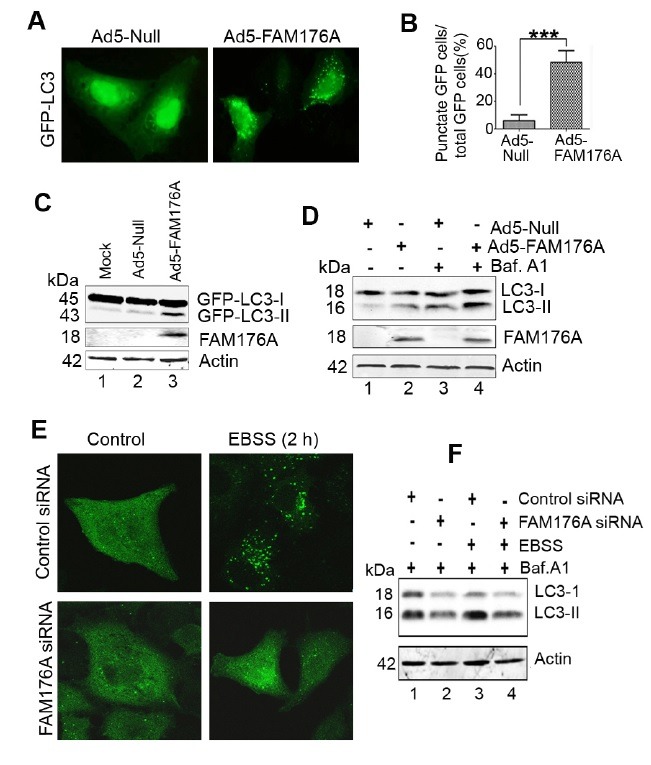
Fig. 2. Ad5-FAM176A induces autophagy in H1299 cells. Knockdown of FAM176A inhibits EBSS-induced autophagy in A549 cells. (A) H1299 cells were infected with either Ad5-FAM176A or Ad5-Null combined with Ad5-GFP-LC3 for 22 h. Fluorescence microscopy was used to observe the punctated distribution of GFP-LC3. Representative fluorescence microphotographs are shown. (B) Statistical analysis was used to test the number of cells with GFP-LC3 punctated distribution. ***P < 0.0001. (C) H1299 cells were treated as in (A). The accumulation of GFP-LC3-II were analyzed by western blot. (D) H1299 cells were infected with Ad5-FAM176A or Ad5-Null at 200 MOI for 20 h, and then treated with 10 nM of bafilomycin A1 for 4 h. The levels of endogenous LC3 and FAM176A expression were analyzed by immunoblotting. (E) A549 cells were transfected with either control siRNA or FAM176A siRNA for 20 h, and then treated with EBSS for 2 h, Fluorescence microscopy was used to observe the punctated distribution of LC3 during autophagy. Representative fluorescence microphotographs are shown. (F) A549 cells were transfected with siRNA as (E), treated with 10 nM of bafilomycin A1 with or without EBSS for 4 h. The levels of endogenous LC3 were analyzed by western blot.