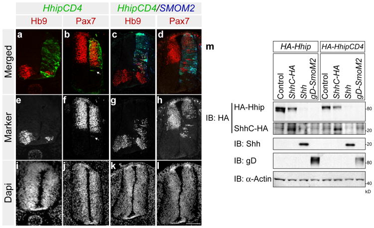
Figure 5. Cell tethered Hhip is not as efficient in inhibiting Shh non-cell autonomously as wild type.
Chick neural tubes were electroporated with pMES-HA-Hhip:CD4 (a, b,), pMES-HA-Hhip: CD4 and pMES-tdTomato-SMOM2 (c, d). HHip:CD4 expressing cells are labelled in green, SmoM2 cells are labelled in blue and HB9 and Pax7 are labelled in red (a–d). Grey scale images of Hb9 and Pax7 (e–h) and corresponding nuclear DAPI satin (i–l). Arrow in b and f indicate that Hhip:CD4 inhibition of Pax7 is cell autonomous. Scale Bar: 50 μm (l). (m) HEK293T cells were co-transfected with Hhip:CD4 and ShhC, Shh, or SmoM2, and Hhip:CD4 was assessed by Western blot.

