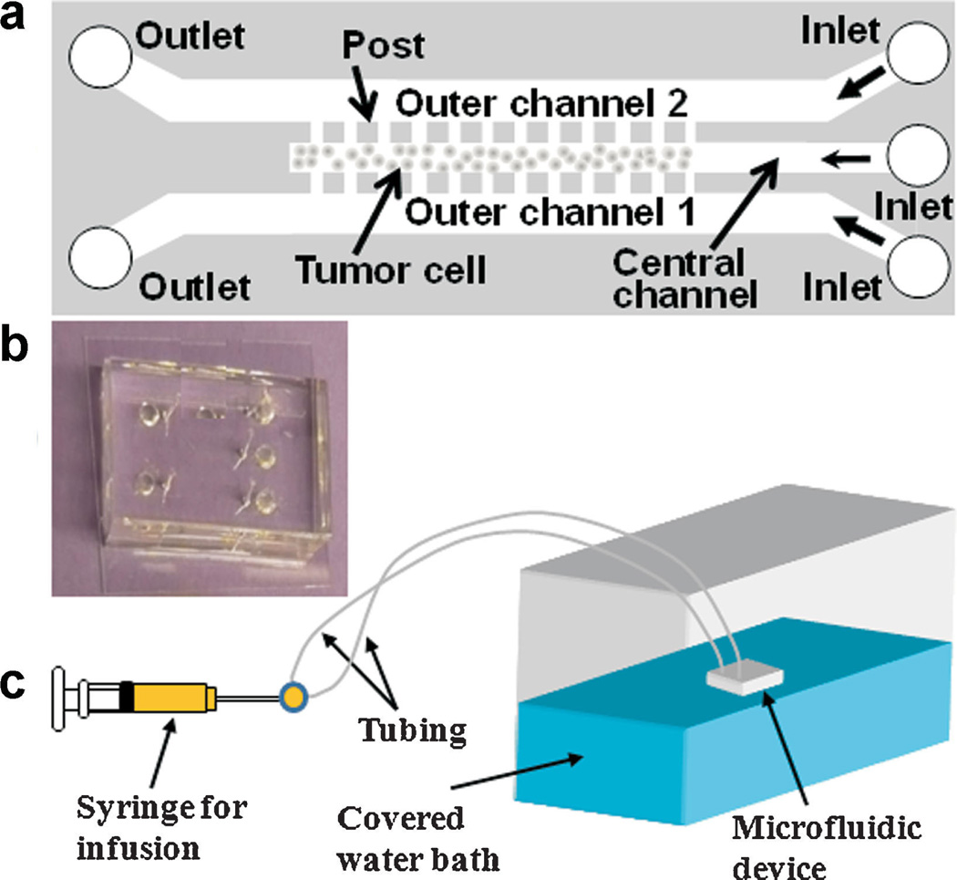
Figure 1.
a: Schematic representation of microfluidic device design for 3D tumor model. The device consisted of three channels that were 0.25 cm in length and 30 µm in height. They were separated by an array of square posts, 50 µm (width) x 30 µm (height), and spaced 4 µm apart. The outer channels were 200 µm wide and the central channel for cell culture was 100 µm wide. b: A photo of the PDMS device. c: Overall setup of the perfusion system for tumor cell culture in the device. The water bath was maintained at 37°C. [Color figure can be seen in the online version of this article, available at http://wileyonlinelibrary.com/bit]

