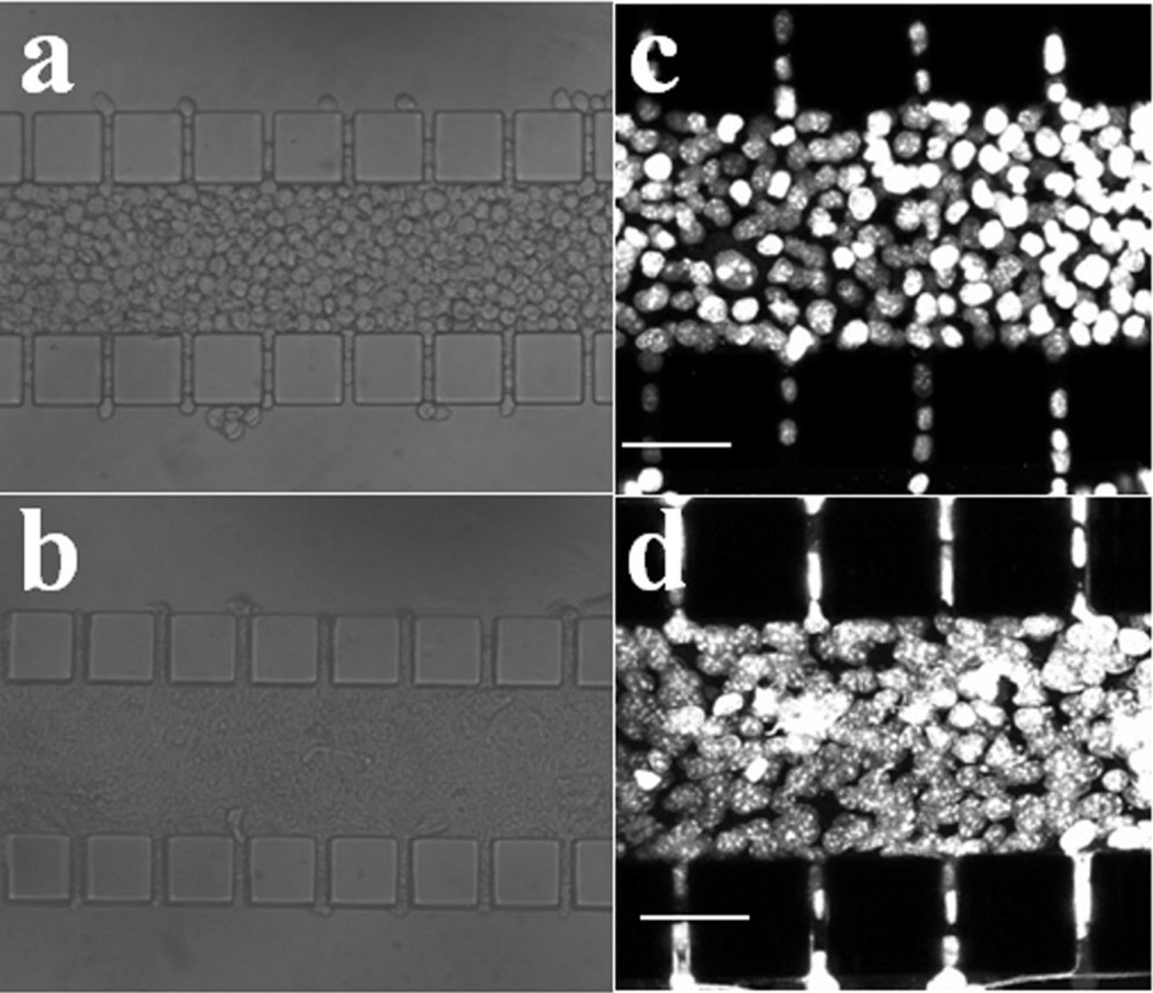
Figure 4.
Images of B16.F10 cells in microfluidic device taken immediately after loading (a,c) and at 12-h post cell loading (b,d). Panels (a) and (b) are images of cell compartment taken under a regular light microscope with trans-illumination; panels (c) and (d) show stacked 2-µm optical slices of the device containing tumor cells with their nuclei labeled with DAPI. These images were taken under a confocal microscope and used for counting cell numbers.

