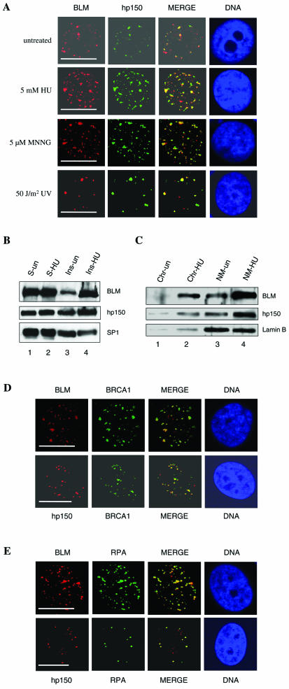
FIG. 3.
BLM and hp150 colocalize within distinct nuclear foci in response to DNA-damaging signals. (A) Indirect immunofluorescence of BLM (red) and hp150 (green) in untreated and HU-, MNNG-, and UV-treated HeLa cells. Representative pictures are shown for a portion of cells analyzed. For details, see Table 1. In the merged image, colocalization is seen as yellow. The panel labeled DNA indicates the position of the nucleus, as judged by DAPI staining. Bars, ∼10 μm. (B) The amount of BLM and hp150 increases after HU treatment in the insoluble fraction of nuclear proteins. Untreated (lanes 1 and 3)and HU-treated (lanes 2 and 4) HeLa nuclear proteins were resolved into soluble (lanes 1 and 2) and insoluble (lanes 3 and 4) fractions, and these were subjected to SDS-PAGE followed by Western blotting for BLM (top panel), hp150 (middle panel), and SP1 (bottom panel [loading control]). (C) The increased amount of BLM and hp150 is mainly associated with the nuclear matrix. After DNase 1 digestion, the insoluble nuclear fraction was separated into two fractions: chromatin (lanes 1 and 2) and nuclear matrix (lanes 3 and 4). Lanes 1 and 3 contain untreated HeLa cells; lanes 2 and 4 contain HU-treated HeLa cells. Lamin B is shown as a fractionation and loading control. (D) BLM and hp150 colocalize with BRCA1 after HU treatment of HeLa cells. Red foci represent either BLM or hp150, and green foci reveal BRCA1 staining. DNA is revealed by DAPI staining. Bars, ∼10 μm. (E) BLM and hp150 colocalize with RPA after HU treatment of HeLa cells. Red represents either BLM or hp150, as indicated, and green indicates RPA protein. DNA indicates the position of nucleus. Bars, ∼10 μm. See Results and Discussion for details.

