FIG. 1.
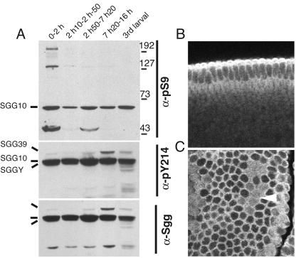
Phosphorylation and subcellular localization of Sgg proteins in embryos. (A) Phosphorylation changes on Sgg during embryogenesis detected by immunoblots of whole extracts by using the indicated phosphorylation state-specific or general antibodies directed against Sgg. Sgg splice forms are labeled together with a molecular weight scale. Other detected bands are unrelated to Sgg. The slightly higher pS9-SGG10 contents in lanes 1, 3, and 5 correlate with embryonic cell division stages: 0 to 2 h (0-2 h), dividing preblastoderm embryos; 2 h 10 min to 2 h 50 min (2 h10-2 h-50), nondividing cellular blastoderm embryos; 2 h 50 min to 7 h 20 min (2 h50-7 h20), germ band elongation stage embryos comprising the last three division cycles; 7 h 20 min to 16 h (7 h20-16 h), organogenesis stages; 3rd larval, third larval instar. A high pY214/general Sgg ratio is observed in embryos compared to cl-8 cultured cells (Fig. 2). (B and C) Immunofluorescent stainings of embryos with the pan-Sgg/GSK-3 antibody MAb 4G1E11. Anterior is left and ventral is down. (B) A transversal confocal section of a stage 5 cellular blastoderm is shown. The staining is excluded from the nuclei and increased in the apical cortex, matching the location proteins required for biogenesis of the zonula adherens. (C) A section through a stage 8 procephalon is shown with staining in the cell cortex. The distribution of Sgg is reorganized in dividing cells of mitotic domains (arrowhead), revealing increased cytoplasmic Sgg.

