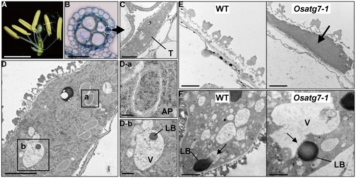
FIGURE 1.
Rice autophagy-defective mutants exhibit sporophytic male sterility, and autophagic degradation within tapetal cells is essential for postmeiotic anther development. (A) Anthers in the wild-type at the uninucleate stage. Scale bar: 3 mm. (B) Transverse sections of wild-type anthers stained with hematoxylin at the uninucleate stage. Scale bar: 50 μm. (C,D) Autophagosome-like structures and lipid bodies enclosed within the vacuoles detected in postmeiotic tapetum cells during pollen development are depicted. Scale bars: 5 μm (C,D) and 1 μm (D-a,b). (E) Potential role of autophagy in tapetal degradation and programmed cell death in rice. Tapetal ultrastructure of the wild-type and the Osatg7-1 mutant at the flowering stage. Scale bar: 1 μm. Arrows indicate the tapetal cell layers. (F) Lipid bodies directly fuse with the vacuoles at the uninucleate stage in the rice tapetum. Scale bar: 1 μm. AP, autophagosome; LB, lipid body; V, vacuole; T, tapetum. Arrows indicate the vacuoles fused with lipid bodies in the wild-type (WT). Similar structure was not observed in the Osatg7-1 mutant.

