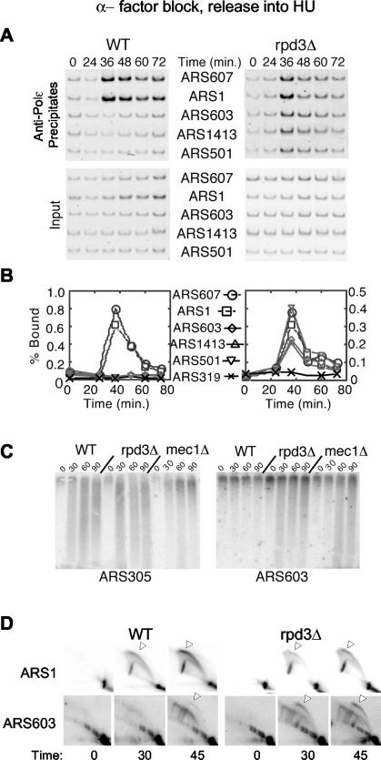
FIG. 3.
Late origins escape checkpoint inhibition in rpd3Δ cells treated with HU. Wild-type (OAy618) and rpd3Δ (OAy781) cells (A and B) or wild-type (OAy470), rpd3Δ (JAy22), and mec1Δ (DGy159) cells (C and D) were synchronized in late G1 with α-factor at 23°C and released into S phase in the presence of 200 mM HU at 23°C. Samples were collected at the indicated intervals. DNA Polɛ association with origins was analyzed by ChIP (A) and quantified (B) as described in the Fig. 1 legend. (C) Nascent DNA strand analysis of ARS305 and ARS603. (D) Replication structures detected by 2-D gel analysis.

