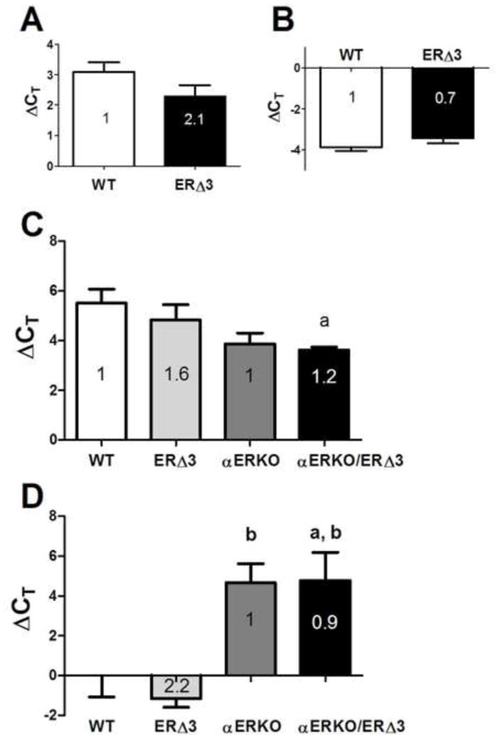
Figure 4. Estrogen responsive genes in the uterus are not repressed by ERΔ3.

Uterine total RNA obtained from 3-month-old female mice in estrus were analyzed for each genotype. Relative mRNA expression was determined by real-time RT-PCR for the progesterone receptor (Pgr), panels A and C, and lactoferrin (Ltf) genes, panels B and D, with normalization to the cyclophilin A (Ppia) gene. In panels A and B, the average ΔCT values are displayed for WT (FVB/N, n=8) and ERΔ3 (line F, FVB/N strain) mice (n=8), with the fold difference determined by the 2−ΔΔCt method indicated within each bar: A. uterine progesterone receptor (Pgr) expression; B. uterine lactoferrin (Ltf) expression. Expression levels in the ERΔ3 mice were not significantly different than in WT uteri (p>0.05, Mann Whitney test). In panels C and D, uteri from intact female mice with a mixed background strain obtained by crossbreeding ERΔ3 (line F, FVB/N strain) and αERKO (C57BL/6 strain) were analyzed. The average ΔCT values for the 4 genotypes, including WT (n=4), ERΔ3 (hemizygous, +/−; n=4), αERKO (homozygous for the ERα disruption, −/−; n=4), and αERKO/ERΔ3 (−/− and +/−, respectively; n=6) are depicted. The fold difference of ERΔ3 relative to WT mice (1.0) and αERKO/ERΔ3 relative to αERKO determined by the 2−ΔΔCt method are indicated within each bar. C. Uterine progesterone receptor (Pgr) expression was significant (p=0.025, 1-way ANOVA) with a significant difference between WT and αERKO/ERΔ3 mice (Tukey’s test). Fold differences relative to WT (1) are 3.1 for αERKO and 3.7 for αERKO/ERΔ3 and relative to ERΔ3 (1) are 1.9 for αERKO and 2.3 for αERKO/ERΔ3. D. Uterine lactoferrin (Ltf) levels were significant (p=0.0042, 1-way ANOVA). The negative ΔCT levels reflect higher Ltf expression compared to the normalizing Ppia gene. Fold differences relative to WT (1) are 0.04 for both αERKO and αERKO/ERΔ3 and to ERΔ3 (1) are 0.2 for the two αERKO genotypes. Genotype comparisons for panels C and D were analyzed by Tukey’s test: a, p<0.05 compared to WT; b, p<0.05 compared to ERΔ3.
