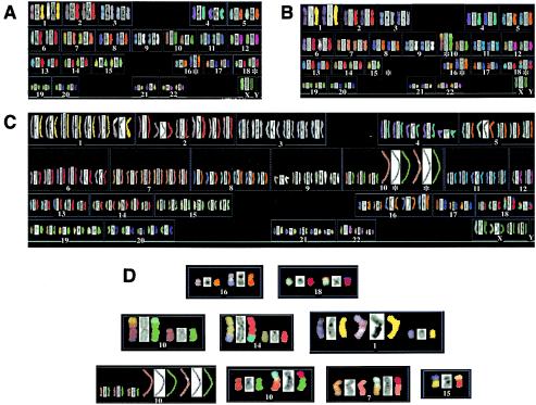
FIG. 5.
Human Ku86-heterozygous cells contain chromosomal abnormalities. SKY analysis of either wild-type cells (A) or two independent Ku86-heterozygous cell lines (B and C). Each chromosome is represented in sets of three corresponding to the actual image, the 4′,6′-diamidino-2-phenylindole (DAPI)-stained image, and the computer-generated, false-color image, respectively. In panel A, the two derivative chromosomes (16 and 18) common to the parental cell line are marked with an asterisk. Panel B shows an example of a Ku86-heterozygous cell that contains—in addition to the parental translocations—a translocation of chromosome 15 to chromosome 10, each of which is also marked with an asterisk. Panel C shows an example of a Ku86-heterozygous cell that has become aneuploid and has sustained an amplification of chromosome 10. Panel D shows individual examples of the parental translocations, as well as some of the abnormalities detected in Ku86-heterozygous cells (see also Table 3).

