FIG. 3.
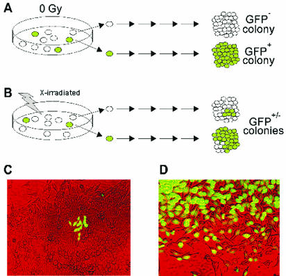
Strategy for detecting delayed instability at the GFP locus. (A) Unirradiated cells are stable and give uniform GFP+ or GFP− colonies. (B) Irradiated cells showing delayed instability involving HR (top) or delayed mutation (bottom). (C and D) Photomicrographs of GFP+/− colonies presumably displaying delayed HR and delayed mutation, respectively.

