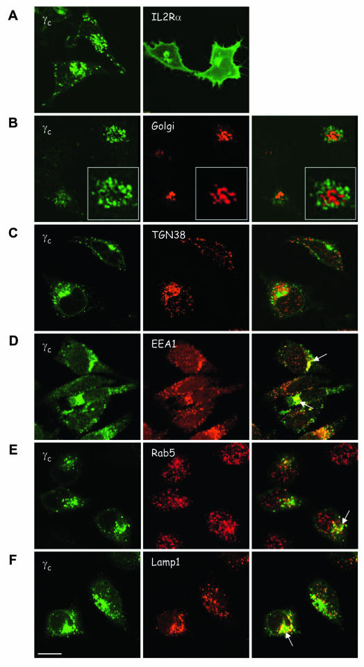
FIG. 2.
Subcellular distribution of γc. HeLa cells were transfected with human γc-GFP (A to F, left panels) or IL-2Rα-GFP (A, right panel). Cells were cotransfected with pEYFP-Golgi marker (B, red) or cells were fixed, permeabilized, and stained with antibodies against various organelles, followed by Alexa-568-coupled secondary antibody (C to F, green). All cells were imaged in confocal mode. (C) TGN38, antibody to the TGN (red); (D) EEA1, antibody to EEA1 (red); (E) anti-Rab5, antibody to early endosomes (red); (F) anti-Lamp1, antibody to lysosomes (red). The white arrows indicate colocalization of γc-GFP with endosomes (D and E) or lysosomes (F). Scale bar, 20 μm.

