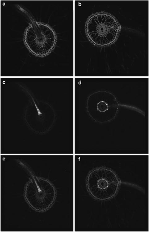
Fig. 1.

Cell type-specific expression of GFP in roots of two GAL4-GFP rice enhancer trap lines as visualized by confocal laser scanning microscopy. Images of root cross sections are presented as fluorescence of the cell wall stain propidium iodide (a, b), GFP (c, d) and overlay of the propidium iodide and GFP fluorescence patterns (e, f). (a, c, e) Enhancer trap line ASG F03 displays bright GFP fluorescence in xylem parenchyma cells and the pattern is most evident in emerging lateral roots. (b, d, f) Enhancer trap line ASU A03 displays bright GFP fluorescence in cortical cells just outside of the endodermis (note that most cortical cells have been replaced by aerenchyma).
