
Figure 5.
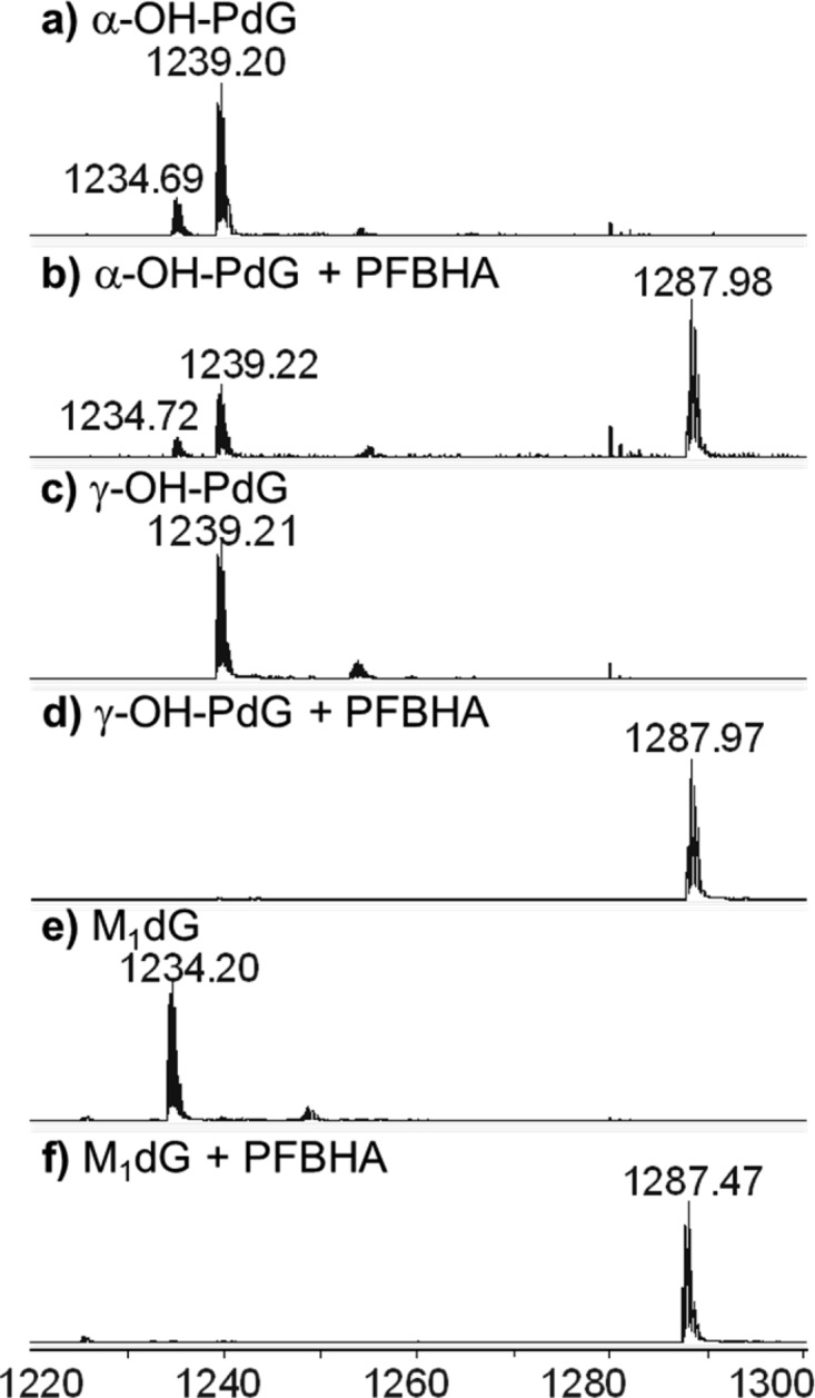
Q-TOF mass spectrometry analysis of the PFBHA trapping reactions of oligonucleotides containing exocyclic guanine lesions. Data represent the −4 charge envelopes, as described in Figure 3. (a) α-OH-PdG; (b) α-OH-PdG + PFBHA; (c) γ-OH-PdG; (d) γ-OH-PdG + PFBHA; (e) M1dG; and (f) M1dG + PFBHA.