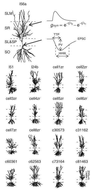
Fig. 1.
Model cell morphologies. Cells have been reoriented so that apical-basal direction is along the y-axis with layer boundaries as indicated. From top (most apical) to bottom (most basal), layers shown are stratum lacunosum-moleculare (SLM), stratum radiatum (SR), stratum lucidum and stratum pyramidale (SL+SP) together, and stratum oriens (SO). Axon segments are not shown. Cell l56a is shown at a larger size to facilitate illustration of an example time course for a synaptic receptor conductance (gsyn) and somatic EPSC along with extracted properties peak value (PV), time-to-peak (TTP), and half-height width (HHW)

