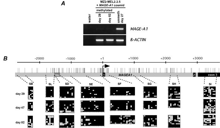
FIG. 2.
Lack of spontaneous demethylating activity targeted to the 5′ region of MAGE-A1 in cells that can express the gene. A cosmid carrying the MAGE-A1 gene within a 42-kb genomic insert was methylated in vitro prior to transfection into MZ2-MEL2.2.5 cells. (A) Expression of MAGE-A1 in these transfectants was analyzed by RT-PCR at two time points after transfection (days 29 and 82) and compared with that in MZ2-MEL2.2.5 cells transfected with the unmethylated MAGE-A1 cosmid (unmeth). (B) Bisulfite sequencing was used to test the methylation state of the in vitro-methylated MAGE-A1 transgene at different time points after transfection. The MAGE-A1 segments that were analyzed and the bisulfite sequencing results are represented as in Fig. 1.

