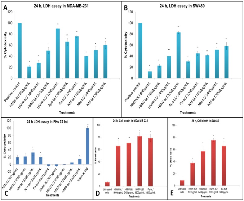
Figure 3. Cytotoxic effects of HMW-bLf.
A B and C represent the cellular cytotoxicity measured by LDH release assay induced by HMW-bLf in a concentration dependent manner, in MDA-MB-231 (human breast carcinoma) SW480 (human colorectal adenocarcinoma) and FHs 74 Int (normal intestinal cells) cells. D and E show the cell death (mortality count) as measured by Flow cytometry using propidium iodide staining (* p<0.05 and ** p<0.01). Other forms of bLf were used for comparison.

