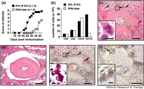
Figure 1.

Accelerated collagen-induced arthritis (CIA) in interferon-γ receptor knock-out (IFN-γR KO) mice is accompanied by CD11b+ splenocyte expansion and osteoclast formation in the joints. Mice were immunised with collagen type II in complete Freund's adjuvant. (a) Accelerated disease onset and more severe arthritic scores in IFN-γR KO mice than in wild-type mice. (b) Expansion of the CD11b+ splenocyte population in IFN-γR KO mice 27 days after immunisation. Splenocytes were obtained from three mice, counted and then pooled for flow cytometric analysis; numbers indicated are per spleen. (c,d) Haematoxylin staining on paraffin sections of the joints on day 27 after immunisation, showing bone erosion and multinucleated giant cells in IFN-γR KO mice (arrows in (c) and detail in inset). (d) Section of wild-type mouse joint, showing normal histological appearance. (e) Tartrate-resistant acid phosphatase (TRAP) staining on paraffin sections of the joint of IFN-γR KO mice, showing multinucleated giant cells (arrows; detail in inset) staining positive for TRAP. TRAP+ multinucleated cells (three or more nuclei) can be considered to be osteoclasts. (f) CD11b+ cells in CIA. Staining with anti-mouse CD11b antibody demonstrating the presence of both CD11b+ (brown) mononuclear cells and multinuclear osteoclast-like cells (arrow; enlarged in inset). The section was counterstained with haematoxylin. Sections that were stained with an isotype control antibody revealed no positive staining (not shown). Bars in the pictures and insets represent, respectively, 100 μm and 10 μm.
