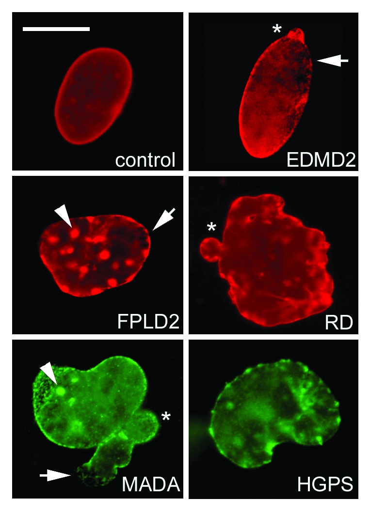
Figure 3. Altered nuclear shape in laminopathies. Representative control, EDMD2, FPLD2, and RD nuclei labeled using anti-lamin A/C antibody (Santa Cruz Sc-6215) are shown in the upper rows, OCT-1 (Santa-Cruz) and farnesylated prelamin A staining (Diatheva 1188–2) of a MADA and HGPS nucleus, respectively, are shown in the lower row. Honeycomb structures, characteristic of EDMD2 (and EDMD1) nuclei are also observed in FPLD2 and MADA (arrows). Lamin A/C aggregates (also labeled by SUN1 and prelamin A) are observed in FPLD2 and MADA (arrowheads), nucler envelope blebs (asterisk) are found in most laminopathies. Bar, 10 μm.
