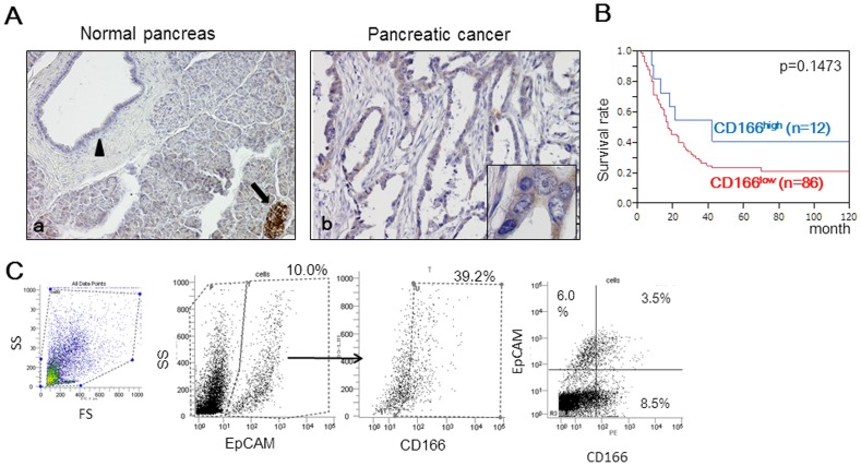
Figure 1. CD166 expression in human pancreatic tissues.
(A) Immunohistochemical staining for CD166 was performed using resected pancreatic tissues. (a) In the normal pancreas, CD166 was expressed strongly in the membrane of islet cells (black arrows) and weakly in normal pancreatic ductal cells (black arrowhead). (b) In some pancreatic cancer tissues, cancer cells were positive for CD166. Original magnification: 200×. Insets: 600×. (B) Kaplan-Meier survival analysis revealed that the intensity of CD166 expression in pancreatic cancer was not correlated with prognosis (p = 0.1473). (C) Flow cytometric analysis of CD166 expression in cells separated based on EpCAM expression. The positive expression rate (%) is indicated for each marker.

