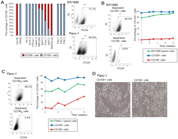
Figure 2. Analysis of CD166 expression in human pancreatic cancer cell lines.
(A) CD166 positivity rates in two normal pancreatic duct epithelial cell lines, pancreatic cancer cell lines, and pancreatic stellate cells (PSCs). (B, C) SW1990 (B) and Panc-1 (C) cells (parental cells) were separated based on CD166 expression (CD166+ and CD166-) by an AutoMACS PRO separator. Changes in CD166 expression in parental and CD166+/− subpopulations were monitored frequently by flow cytometry over 6 weeks. (D) Morphology of Panc-1 cells separated based on CD166 expression. Original magnification: 40×.

