Figure 6.

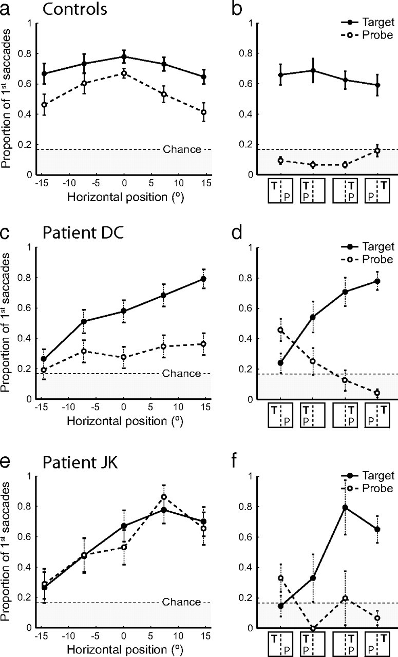
Orienting to separately and simultaneously presented targets and probes. a, Proportion of first saccades directed toward the target letter on target-only trials (solid line) and toward the probe letter on probe-only trials (dashed line) by healthy controls, as a function of the horizontal position of the target/probe. b, Proportion of first saccades directed toward the target letter (solid line) and probe letter (dashed line) by controls on target-plus-probe trials, as a function of the horizontal locations of target (T) and probe (P) to left or right of subject midline. Error bars indicate SEM. c, d, Corresponding results for patient D.C., a neglect patient with a posterior parietal lesion, plotted as in a and b. Note that both target (solid line) and probe responses (dashed line) display a gradient of impairment from normal performance at far right (compare c, a), falling to chance performance at far left. Error bars indicate SE of the binomial probability estimate. e, f, Corresponding results for patient J.K., a frontal neglect patient without parietal damage. Note that both target and probe responses display a horizontal gradient of impairment as in parietal neglect. Additionally, orienting to ipsilesional (rightward) probes in the probe-only condition is enhanced in comparison to controls (compare dashed lines in e, a).
