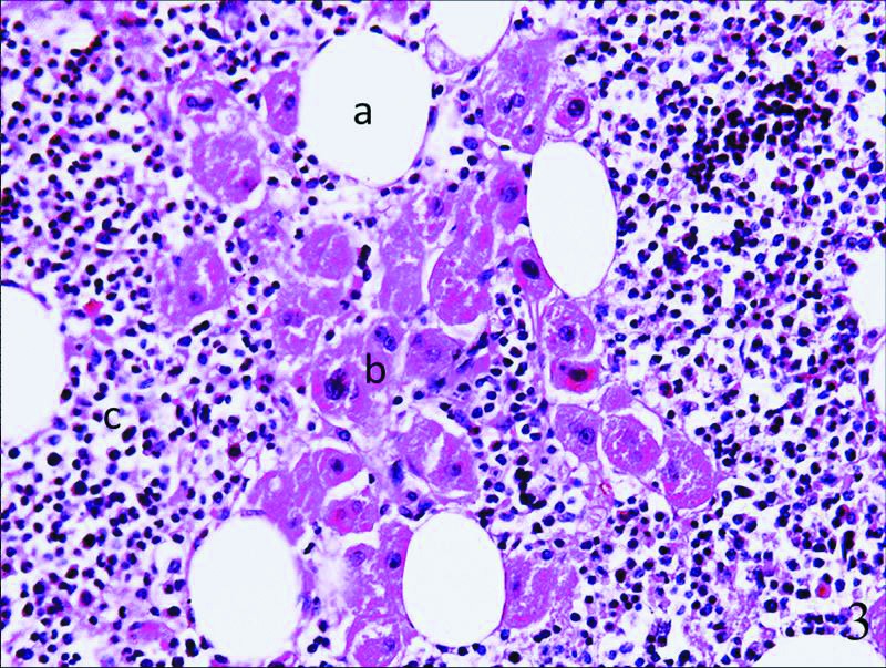
Fig. 3.

At the microscopic level, the lesions were composed of mature adipose tissue (a), scattered islands of hematopoietic cells (b), and hyperplasia of the adrenal cortex (zona reticularis) (c), with adipose tissue (a) and hematopoietic elements (b) being among the adrenal tissues.
