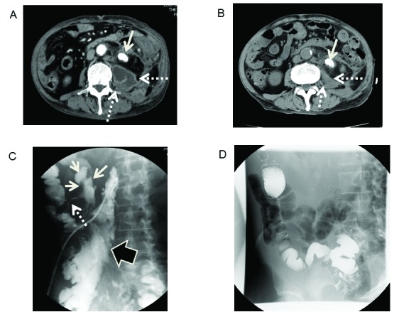
Fig. 1.

(A) Contrast-enhanced computed tomography (CT) revealed the left ureter stone in the uretero-pelvic junction (solid arrow), and the perinephric abscess (dotted arrow). (B) Plain CT revealed abscess shrinkage for 6 months after treatment (dotted arrow). (C) Antegrade contrast radiography of an abscess (black arrow) revealed a fistula into the descending colon (dotted arrow), and the left hydronephrosis (solid arrow). (D) Barium enema of the colon revealed no apparent communication to the abscess.
