
Figure 2.
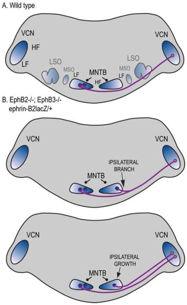
Altered brainstem projections in mice with mutations that reduce EphB signaling. A. Normal pathway from AVCN and PVCN (indicated as VCN) to MNTB in wild type mice. Nearly all of the projections are contralateral, terminating in a calyx of Held in the appropriate frequency region. Blue color gradients refer to frequency axis; dark blue represents high frequencies. LSO, lateral superior olive; MSO, medial superior olive. B. Mutations that block reverse signaling through ephrin-B receptors result in a significant number of ipsilateral calyceal projections (Hsieh et al., 2010). These ipsilateral projections in most cases appear as branches from contralaterally projecting VCN axons. In some cases, ipsilateral projections grow directly to the MNTB in the absence of a contralaterally projecting branch. For clarity, MSO and LSO are omitted; these nuclei have not been studied in the context of Eph signaling.