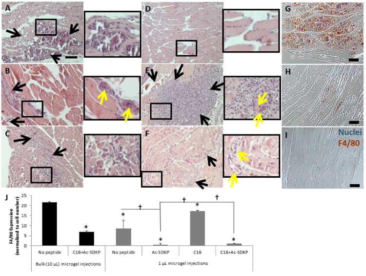
Figure 2. Macrophage infiltration with injectable microgels in ischemic muscle.

Sections of adductor muscle tissue adjacent to peptide-loaded injectable polymer microgels were stained with hematoxylin and eosin (H&E, A-F) or rat anti-mouse biotinylated F4/80 antibodies (a macrophage marker), as visualized by brown color in images (G-I). Nuclei were counterstained blue with hemalum. Scale bar =100 μm. Black rectangles indicate magnified area. Black arrows indicate inflammatory infiltration. Yellow arrows indicate blood vessels. Bulk 10 μL injection of microgels without peptide resulted in a high level in inflammatory infiltration (A, G), which was decreased with the co-treatment of C16 and Ac-SDKP peptides (B, H). Multiple 1 μL microgel injections (C) decreased the inflammatory response compared to the bulk injection (A). When delivered with multiple microgel injections, anti-inflammatory Ac-SDKP (D) minimized the inflammatory response, while C16 (E) increased the inflammatory response. The co-treatment (F, I) prevented inflammatory exacerbation while still forming blood vessels. (J) F4/80 staining was quantified by calculating the area of positively stained pixels divided by the total number of cells per image as measured by hematoxylin nuclear stain. Data are means ± SEM from four mice per condition. *p<0.05 vs. bulk injection with no peptide; †p<0.05 vs. groups connected by lines (Tukeys' Range Test).
