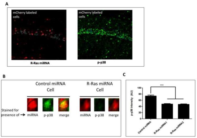
Figure 3. Knockdown of R-Ras in CA1 attenuates p38 MAPK activation after HFS-LTP stimulation.
Hippocampal slices prepared from mice injected with viruses expressing R-Ras miRNA1 or R-Ras miRNA2 into CA1 were stimulated for HFS-LTP and processed for immunostaining of phosphorylated p38. A. Representative image of CA1 pyramidal neurons positive for miRNA (mCherry) (left) traced and overlayed onto the CA1 layer of p-p38-labeled cells. Fluorescent intensity of p-p38 was quantified for each R-Ras miRNA positive cell and compared to the population of non-infected cells. B. Magnified region of A showing examples of p-p38 in both uninfected cells and cells infected with R-Ras miRNA expressing virus in the same brain slice. C. Quantification of p-p38 fluorescent intensity in R-Ras miRNA1− or R-Ras miRNA2− positive cells compared to virus-negative cells (One-Way ANOVA, F 2,732 = 199, p < 0.0001, Bonferroni post-hoc: R-Ras miRNA1+ vs R-Ras miRNA−, t= 15.93; p < 0.05, R-Ras miRNA2+ vs R-Ras miRNA−, t = 18.04, p < 0.05.

