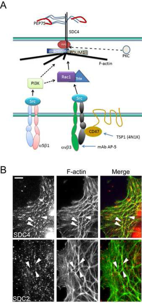
Figure 1. Schematic of CLAN components and integrin-mediated signaling pathways involved.
A) Syndecan-4 (SDC4), α-actinin, PIP2 and PDLIM1 have previously been shown to localize within the vertices of interconnecting F-actin bundles in CLANs where we propose they may form a molecular signaling complex or “vertisome” [19, 28]. Other known CLAN components not depicted include filamins A and B, myosin and tropomysosin [19, 20, 22, 28].The formation of this structure has been shown to involve distinct β1 and β3 integrin pathways that utilize PI3-kinase (PI3K) and CD47/Rac1/Trio respectively [31]. PI3K may activate Trio, or it may converge with the Rac/Trio pathway downstream of Rac1 (dotted line). The formation of this CLAN structure can be triggered by PEP75 (red curved line) which is a peptide from laminin 5 that can bind syndecan-4. It can also be triggered by an activating antibody to αvβ3 integrin (mAb AP-5) or a peptide from thrombospondin-1 (TSP1) called 4N1K [31]. PKCs may also activate other signaling molecules that regulate CLAN formation. B) Syndecan-4 (SDC4) is an integral part of the CLAN structure while syndecan-2 (SDC2) is not. The merged image shows that syndecan-4 localizes specifically to the vertices of the CLAN structure. This localization is not observed for syndecan-2. Arrowheads show location of CLAN vertices obvserved with F-actin labeling. Scale bar = 5 µm.

