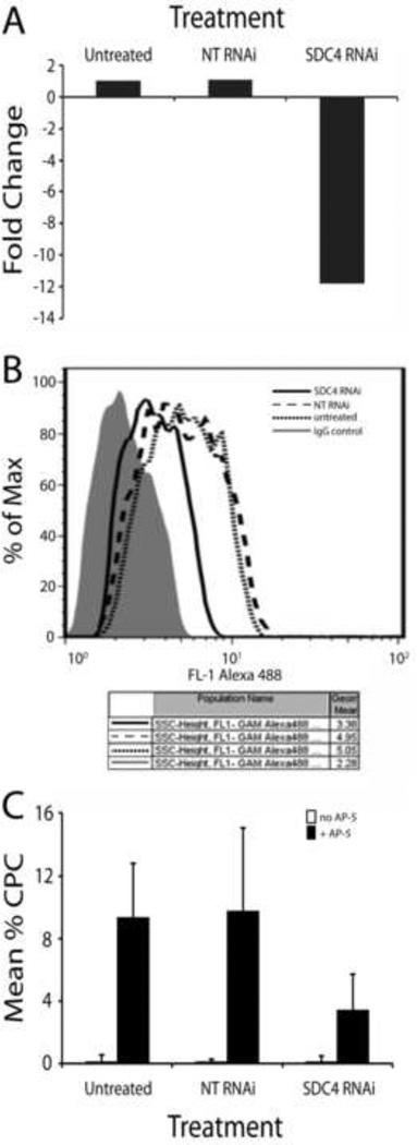
Figure 2. Loss of syndecan-4 reduces β3 integrin-mediated CLAN formation.
A) N27TM-3 HTM cells were left untreated or transfected with a non-targeting (NT) RNAi or syndecan-4 (SDC4) RNAi. 72 hours post-transfection, RNA was extracted and analyzed for loss of RNA expression using real-time PCR. Cells were processed as described in Materials and Methods. Data were normalized to GAPDH and the fold change compared to untreated cells was determined. B) FACS analysis was performed on HTM cells transfected with NT or SDC4 RNAi 72 hours post-transfection to determine cell surface expression of SDC4. Cells were processed as described in Materials and Methods. C) Untreated cells or cells treated with NT or SDC4 RNAi were re-plated onto fibronectin-coated coverslips 72 hours post-transfection in the absence or presence of the β3 integrin-activating antibody AP-5. Three hours later the cells were fixed and labeled with phalloidin. The percentage of CLAN-positive cells (CPCs) is shown as the mean ± the SD; n ranged from 1295-1582 cells for each treatment group. For all 3 treatment groups, the cells treated with mAb AP-5 showed increased CLAN formation over the corresponding untreated cells (P < 0.01). For AP-5-treated cells, SDC4 RNAi significantly reduced CLAN formation over untransfected cells or cells transfected with non-targeting RNAi (P < 0.01).

