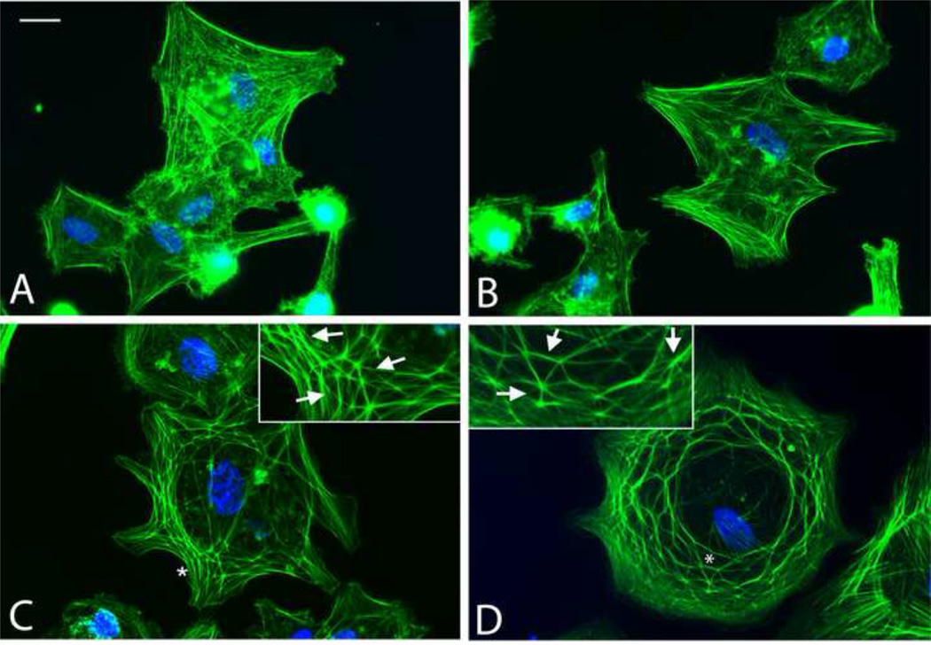
Figure 5. Comparison of HTM cells treated with PepCon, PEP75 or mAb AP-5.
N27TM-2 HTM cells were plated onto fibronectin-coated coverslips and allowed to spread for 3 hours prior to fixation and labeling with Alexa 488-conjugated phalloidin and Hoechst 33342. A) untreated cells; B) cells treated with 60 µg/mL PEPcon; C) cells treated with 60 µg/mL PEP75 and D) cells treated with mAb AP-5. Insets = enlarged areas of cells with CLANs; asterisks = areas in panels C & D enlarged in insets. Scale bar = 20µm.

