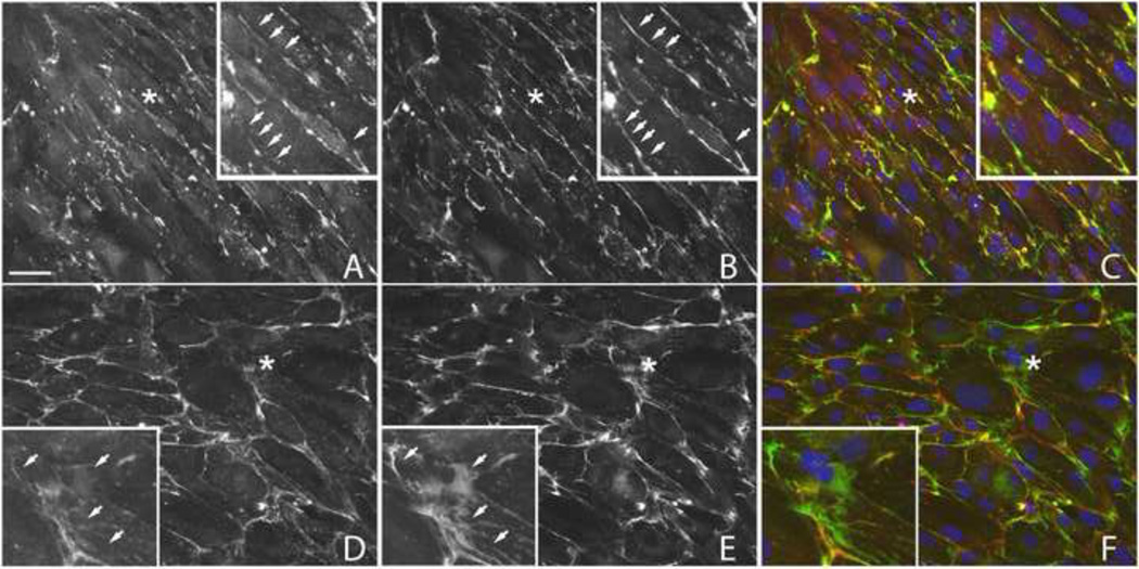
Figure 7. Localization of laminin 5 and laminin 1 in HTM cells treated with or without dexamethasone.
Confluent monolayers of N27TM-2 cells were treated for 7 days with 0.1% ethanol (A–C) or 500 nM DEX (D–F) prior to fixation and labeling with either mouse mAb P3H9-2 against laminin 5 (A, D; red channel) or a rabbit pAb against laminin 1 (B, E; green channel). DEX increased the labeling for both laminins, however, the labeling patterns for laminin 5 and laminin 1 did not completely overlap. Merged images (C, F) are also shown. Similar results were obtained with the N17TM-2 HTM cell strain (data not shown). Asterisks = areas enlarged within insets; arrows = regions where laminin 1 and laminin 5 labeling show incomplete co-localization. Scale bar = 50 µm.

