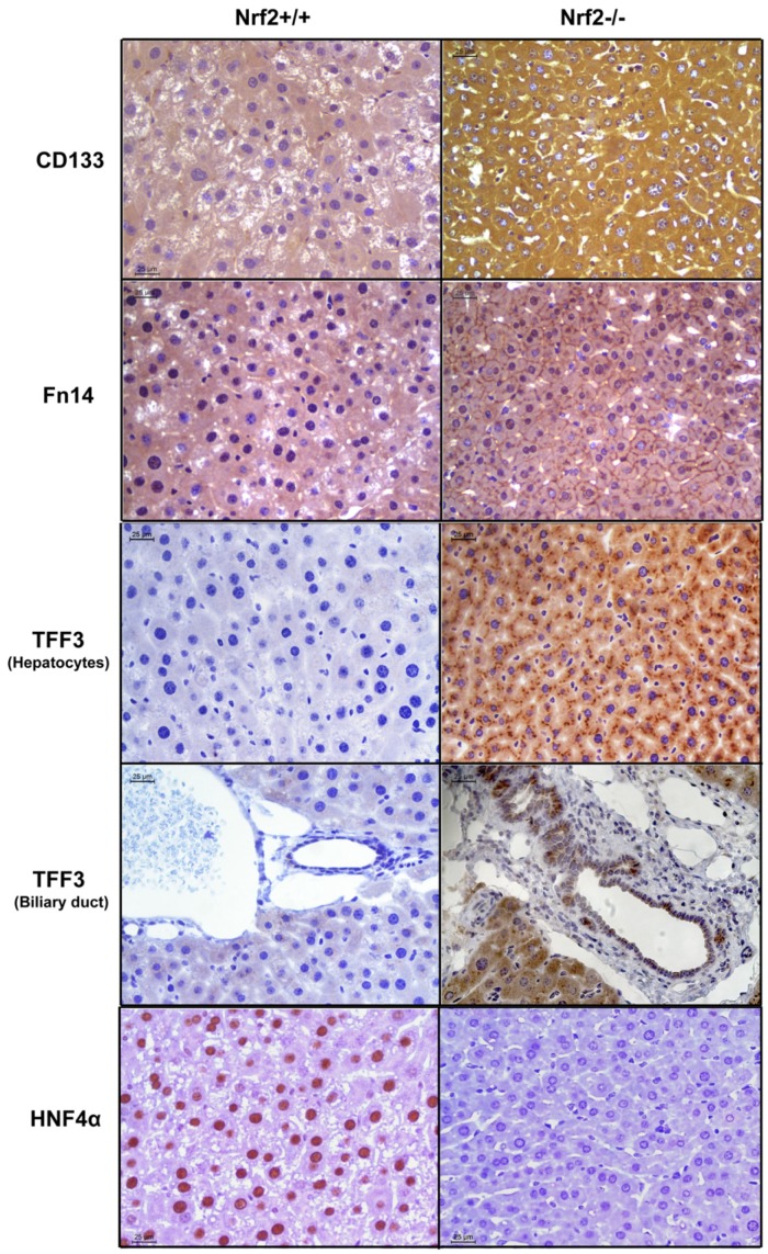
Figure 4. Immunohistochemical analysis of CD133, Fn14, TFF3, and HNF4α in regenerating livers of Nrf2+/+ and Nrf2−/− mice.
Liver sections were prepared from the livers isolated at 60 h after PH from three mice per genotype. All liver sections were subjected to immunostaining with primary antibodies against CD133, Fn14, TFF3, or HNF4α. Representative immunohistochemically stained liver sections are shown.

