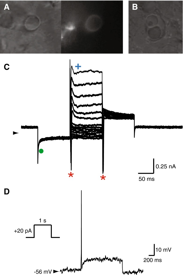
FIG. 5.
Recordings from a calyx primary afferent terminal in 15 WG fetal vestibular neuroepithelium. A Infrared DIC image shows a calyx terminal (viewed from above) that appears as a ring. Fluorescent image shows a similar “halo” after filling the terminal with Alexa-594 dye during recording. Note: hollow appearance of halo. B Complex calyces were also observed at this age. C Voltage clamp recording from the calyx primary afferent terminal shows activation of potential IK,L (filled green circle), Na+ (red asterisks), and K+ (blue cross) whole-cell currents in response to voltage activation protocol (as shown in Fig. 2A inset). D Current clamp recording shows a single overshooting action potential generated by a depolarizing current step injection (inset) in the same calyx primary afferent terminal.

