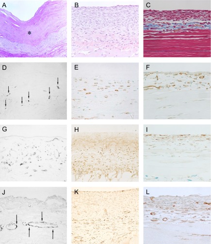
Fig. 2. .
Peritonea from long-term peritoneal dialysis patients (A), (D), (G), and (J). Hematoxylin and eosin staining of peritoneal tissue from a patient presenting ultrafiltration loss. Mesothelial cells were mostly detached, and peritoneal tissue was markedly thickened, by the irregular proliferation of collagen fibers (×100). * Indicates hyalinous changes of collagen fibers (A). Many cells positive for CD68, a macrophage marker (arrow) were present in the area thickened with the collagen fibers (×100) (D). The α-smooth muscle actin (SMA) was abundantly expressed in the thickened submesothelial compact zone (×400) (G). Blood vessels (arrow) were observed in the lower area, in the submesothelial compact zone (×100) (J). These figures are extracted from Shioshita, et al. 2000 [57] with permission. The peritoneal tissues from the mouse CG model are shown in (B), (E), (H), and (K). The injection of CG induced a significant thickening of the peritoneum within 3 weeks (×100) (B). F4/80-positive macrophages were found in the submesothelial compact zone (×400) (E). Note the presence of a large number of α-SMA-positive cells in the submesothelial compact zone (×200) (H). Numerous vessels were positively stained for CD31 (×200) (K). These figures are extracted from Arai et al. 2011 [2] and Nakazawa et al. 2013 [44] with permission. The peritoneal tissues obtained from the MGO-induced peritoneal fibrosis model (C), (F), (I), and (L). As shown in masson-trichrome staining, the injection of MGO induced a significant thickening of the peritoneum (×200) (C). F4/80-positive cells in the markedly thickened peritoneal tissues (×200) (F). Note the presence of numerous α-SMA-expressing cells in the thickened submesothelial compact zone (×200) (I). Numerous vessels that stained positive for CD31 appeared in the lower submesothelial compact zone (×200) (L). These figures are extracted from Kitamura et al. 2012 [32] with permission.

