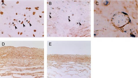
Fig. 3. .

Note the abundant expression of proliferating cell nuclear antigen (PCNA)-positive cells among the vascular endothelial cells in the CG-induced peritoneal fibrosis model (arrowheads) (×400) (A). Administration of TNP-470 to the CG-induced peritoneal fibrosis model decreased the number of PCNA-positive cells in the submesothelial area (B). Note that CD31-positive cells were also positive for PCNA (×800). Double staining for CD31 (brown) and PCNA (blue) in the same section (C). Immunohistochemistry for collagen type III showed that the administration of TNP-470 suppressed significantly the progression of peritoneal thickening (E) compared to that of the untreated mouse (×200) (D). These figures are extracted from Yoshio et al. 2004 [62] with permission.
